Introduction
HVAC design for MRI suites is one of the most technically challenging aspects of MRI room construction. The MRI scan room must maintain precise temperature and humidity conditions while managing significant heat loads — all without compromising the integrity of the RF shielding enclosure (Faraday cage).
The fundamental challenge is that every duct penetrating the shielded enclosure creates a potential RF leakage path. The solution is the RF waveguide — a carefully sized hollow tube that allows air to pass through the shield while attenuating RF energy below the frequency of concern. Properly designing, sizing, and installing these waveguides is critical to both shielding effectiveness and patient/equipment climate control.
Temperature & Humidity Requirements
MRI Scan Room
MRI manufacturers specify tight environmental conditions for the scan room to ensure optimal scanner performance and patient comfort:
- Temperature: typically 18–22°C (64–72°F), with some manufacturers specifying a narrower range of 20 ± 1°C. Temperature stability is as important as the absolute value — fluctuations can cause thermal drift in the magnet and electronics.
- Relative humidity: 30–60% RH, non-condensing. High humidity risks condensation on cryogenic components, while low humidity increases static discharge risk near sensitive electronics.
- Air change rate: minimum 6–10 air changes per hour (ACH) is typical for adequate ventilation and temperature uniformity.
Equipment Room
The equipment room housing gradient amplifiers, RF amplifiers, and system electronics has more demanding cooling requirements. These components can generate 20–40 kW of continuous heat during scanning sequences. Equipment room conditions are typically:
- Temperature: 18–24°C (64–75°F), with some gradient amplifiers requiring temperatures below 20°C
- Dedicated cooling: separate HVAC zone from the scan room, often requiring supplemental cooling (split systems or chilled water units) to handle the concentrated heat load
Chiller/Cooling System
Most MRI systems require a dedicated water cooling loop (chiller) for the gradient coils and, in some older systems, the RF amplifier. The chiller is typically located outside the building or in a mechanical room, with chilled water piping running to the equipment room. The chiller capacity ranges from 20–60 kW depending on the MRI model and gradient performance level.
Heat Load Calculation
Accurate heat load calculation is essential for sizing the HVAC system. The MRI scan room heat load includes:
MRI Scanner Heat Dissipation
The MRI system dissipates heat from several sources:
- Cold head compressor: 3–8 kW (operates continuously to maintain helium at cryogenic temperatures)
- Gradient coils: 5–20 kW during scanning (intermittent, varies with scan protocol)
- RF body coil: 2–5 kW during scanning
- Patient: approximately 100 W (sensible heat from a resting adult)
MRI manufacturers provide detailed heat dissipation data for each scanner model in their site planning documentation. The HVAC engineer must use these specific values rather than generic estimates.
Lighting and Equipment
Non-ferromagnetic lighting fixtures, patient monitoring equipment, and any auxiliary devices in the scan room contribute additional heat load, typically 0.5–1.5 kW total.
Building Envelope
Heat gain or loss through the room's walls, floor, and ceiling (transmission load) is typically modest in interior MRI rooms but significant for rooms with exterior walls. The RF shielding layers (metal panels) and any magnetic shielding steel add thermal mass and slight insulation value to the room envelope.
Total Cooling Requirement
A typical 1.5T MRI scan room requires approximately 8–15 kW (2.3–4.3 tons) of cooling capacity. A 3T system with high-performance gradients may require 12–25 kW (3.4–7.1 tons). These values are scan-room only; the equipment room typically requires an additional 15–40 kW of dedicated cooling.
RF Waveguides for HVAC Penetrations
RF waveguides are the engineered solution for passing airflow through the Faraday cage without compromising shielding effectiveness. A waveguide is a hollow tube — typically round or rectangular — that acts as a high-pass filter for electromagnetic energy: frequencies below the cutoff frequency are attenuated exponentially as they travel through the tube, while air passes freely.
Cutoff Frequency
The cutoff frequency of a circular waveguide is determined by its diameter:
where fc is the cutoff frequency, c is the speed of light (3 × 10⁸ m/s), and d is the internal diameter. For RF energy at frequencies below fc, the waveguide provides attenuation of approximately 32 dB per diameter of length.
Sizing for MRI Applications
For the waveguide to attenuate the MRI's operating frequency, its cutoff frequency must be higher than the Larmor frequency. This means the waveguide diameter must be smaller than the cutoff diameter for the frequency of concern:
- 1.5T (63.87 MHz): cutoff diameter approximately 2.76 m — any practical duct size is far below cutoff, providing excellent attenuation. Standard HVAC duct sizes (150–300 mm) work well.
- 3T (127.74 MHz): cutoff diameter approximately 1.38 m — again, standard duct sizes provide ample attenuation margin.
The critical factor is waveguide length. To achieve sufficient attenuation (typically 80+ dB), the waveguide length must be at least 3–4 times its diameter. For a 200 mm (8-inch) diameter waveguide, the minimum length should be 600–800 mm.
Waveguide Construction
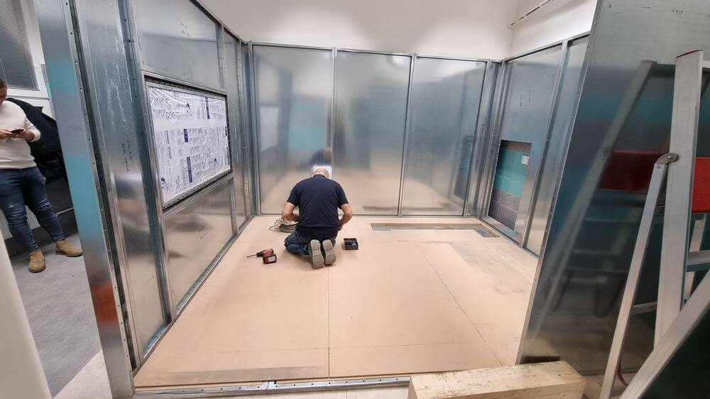
HVAC waveguides for MRI must be constructed from the same type of conductive material as the Faraday cage — typically copper or aluminum tubes or sheet-metal duct with welded or soldered seams. The waveguide must be bonded to the Faraday cage wall with a continuous, electrically conductive seal around its full circumference. Any gap or poor bond at this connection is a common source of SE test failures.
Honeycomb Waveguide Arrays
When larger airflow volumes are needed but waveguide attenuation must be maximized, honeycomb waveguide panels can be used. These consist of an array of small-diameter tubes (typically 10–25 mm) bundled together to form a panel. The small individual tube diameters provide very high cutoff frequencies, while the collective cross-sectional area provides adequate airflow. Honeycomb panels are often used for the main supply and return air penetrations.
Ductwork Layout & Design
Supply Air Distribution
The supply air distribution inside the MRI scan room must provide uniform temperature without creating drafts that could affect patient comfort or cause image artifacts from patient movement. Common approaches include:
- Ceiling diffusers: non-ferromagnetic diffusers (aluminum or plastic) mounted in the ceiling, fed by waveguide penetrations in the ceiling shield. This approach provides good temperature uniformity.
- Perimeter slot diffusers: linear diffusers along wall-ceiling junctions for gentle, draft-free air distribution.
- Underfloor supply: in some configurations, conditioned air is supplied through the floor and returns through ceiling vents, creating a displacement ventilation pattern.
Return Air Path
Return air exits the scan room through separate waveguide penetrations, typically located at the opposite end or side from the supply diffusers to ensure full room circulation. The return air path should not short-circuit the supply air — adequate separation between supply and return points is essential.
Positive Pressure
Maintaining slight positive pressure (2.5–5 Pa) in the MRI scan room relative to adjacent spaces prevents unfiltered air, dust, and contaminants from entering through the RF door seal when the door is opened. This also helps maintain consistent temperature conditions.
Emergency Ventilation & Oxygen Monitoring
In addition to normal HVAC, MRI rooms with superconducting magnets require emergency ventilation systems to address the risk of helium release into the scan room.
Oxygen Depletion Risk
If the magnet quench pipe fails or a slow helium leak occurs, gaseous helium can accumulate in the scan room, displacing oxygen. Helium is lighter than air and accumulates first at ceiling level, but can rapidly fill the room in a major release. Oxygen levels below 19.5% (OSHA threshold) are hazardous; levels below 16% are immediately dangerous to life.
Oxygen Monitoring System
An oxygen depletion sensor (or multiple sensors) must be installed in the MRI scan room, typically at ceiling level where helium accumulates first. The system should alarm at 19.5% O₂ (OSHA action level) and trigger the emergency exhaust ventilation system. Audio and visual alarms must be present both inside and outside the scan room.
Emergency Exhaust Fan
A dedicated emergency exhaust fan, activated by the oxygen monitoring system, rapidly ventilates the scan room in a helium release event. This fan typically provides 15–20 air changes per hour and exhausts to the building exterior. The exhaust duct also penetrates the Faraday cage through a waveguide. The emergency ventilation system must be on emergency power (generator backup) and must be independent of the normal HVAC system.
Compliance
Emergency ventilation requirements are addressed in NFPA 99 (Health Care Facilities Code), the ACR Guidance Document on MR Safe Practices, and individual state building codes. The design must be reviewed and approved by the Authority Having Jurisdiction (AHJ) and the facility's safety officer.
Coordination Between HVAC & Shielding Contractors
Poor coordination between the HVAC contractor and the RF shielding contractor is one of the most common causes of construction delays and SE test failures in MRI projects. Best practices include:
- Joint design review: the shielding contractor, HVAC engineer, and MEP consultant should review waveguide locations, sizes, and connection details together before construction begins.
- Waveguide responsibility: clearly define whether the shielding contractor or the HVAC contractor is responsible for furnishing and installing waveguides. In most projects, the shielding contractor provides the waveguides as part of the Faraday cage package.
- Duct connections: the HVAC contractor connects their ductwork to the exterior face of the waveguide. No standard ductwork (galvanized steel, flexible duct) should penetrate the shielding plane.
- No penetration modifications: once the shielding is installed and tested, no additional HVAC penetrations should be made without consulting the shielding contractor. Unauthorized penetrations will void the shielding warranty and compromise SE.
- Pre-test inspection: both contractors should inspect all waveguide connections before the SE test to verify proper bonding and sealing.
Frequently Asked Questions
What is an RF waveguide in MRI room HVAC?
An RF waveguide is a hollow metal tube (typically copper or aluminum) that allows air to pass through the MRI Faraday cage while blocking RF energy. It works as a high-pass filter: RF signals below the waveguide's cutoff frequency are attenuated exponentially as they travel through the tube, while air flows freely. Waveguides must be properly sized (length at least 3 to 4 times the diameter) and bonded to the Faraday cage with continuous conductive seals.
What temperature does an MRI room need to be?
MRI scan rooms typically require 18 to 22 degrees Celsius (64 to 72 degrees Fahrenheit) with some manufacturers specifying a tighter range of 20 plus or minus 1 degree Celsius. Relative humidity should be maintained at 30 to 60 percent, non-condensing. Temperature stability is critical because fluctuations can cause thermal drift in the magnet and electronics, potentially affecting image quality.
How much cooling does an MRI room need?
A typical 1.5T MRI scan room requires approximately 8 to 15 kW (2.3 to 4.3 tons) of cooling capacity, while a 3T system may require 12 to 25 kW (3.4 to 7.1 tons). The equipment room typically needs an additional 15 to 40 kW of dedicated cooling for gradient amplifiers and electronics. Exact requirements depend on the specific MRI model and are provided in the manufacturer's site planning documentation.
Why do MRI rooms need oxygen sensors?
MRI rooms with superconducting magnets need oxygen sensors because the liquid helium coolant can leak or be released during a magnet quench event. Helium gas displaces oxygen and is odorless and invisible. An oxygen depletion sensor triggers alarms and activates emergency exhaust ventilation when oxygen levels drop below 19.5 percent, protecting patients and staff from asphyxiation risk.
Can standard HVAC ductwork be used in an MRI room?
Standard HVAC ductwork (galvanized steel, flexible duct) cannot penetrate the MRI Faraday cage directly, as it would create RF leakage paths. All duct penetrations through the shielded enclosure must pass through RF waveguides made of the same conductive material as the Faraday cage. Inside the scan room, non-ferromagnetic ductwork and fittings (aluminum or copper) must be used within the 5-gauss fringe field line.