
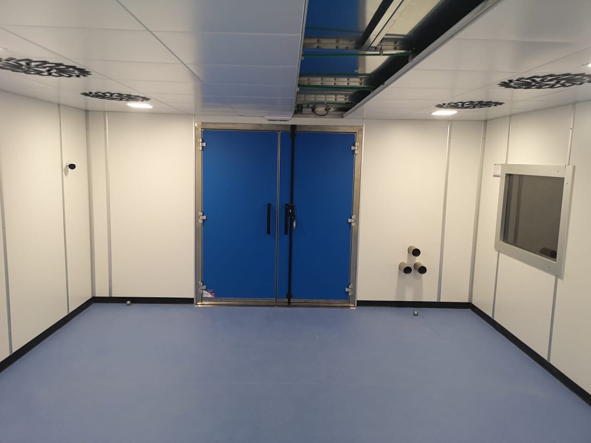
Why veterinary MRI suites need the same shielding as human facilities
Magnetic resonance imaging has become a standard diagnostic tool in veterinary medicine, from university teaching hospitals to private specialty practices. The physics of the scanner is identical whether the patient is a human or an animal: the receiver coils detect extremely weak radio-frequency signals, and any external interference degrades image quality. For this reason, a veterinary MRI suite requires the same RF shielding fundamentals — a properly designed and tested Faraday cage — as any human clinical installation.
What differs is not the shielding physics but the operational context around it. Veterinary facilities handle patients under general anesthesia in nearly every case, work with a wide range of animal sizes, and often integrate the MRI into a building that was not originally designed for medical imaging. These factors shape how the shielded enclosure is designed, where penetrations are placed, and how the room supports the clinical workflow.
Anesthesia and animal handling drive the room layout
Unlike human MRI, where most patients cooperate while awake, veterinary imaging is performed almost entirely under general anesthesia. This has a direct effect on the shielded room design. Anesthesia gas lines, monitoring cables, and ventilator connections all need to pass through the RF enclosure, and each of these penetrations must be handled through properly filtered penetration panels or waveguides so that the integrity of the Faraday cage is preserved.
The room layout must also allow the clinical team to move an anesthetized animal safely onto the scanner table and to position monitoring equipment where it can be observed. Because patient sizes range from small companion animals to large breeds and occasionally equine limbs, the door opening, table access, and clear floor space are planned around the practice's expected caseload. A well-designed veterinary suite keeps anesthesia and monitoring equipment accessible while maintaining the sealed boundary of the shielded enclosure.
Retrofitting existing veterinary buildings
Many veterinary MRI installations go into buildings that were originally constructed for general practice or surgery rather than advanced imaging. This makes the shielded room a retrofit project rather than new construction, which introduces constraints on ceiling height, floor loading, and access routes for delivering the scanner and shielding materials.
The modular Faraday cage approach is often well suited to these situations. A modular shielded enclosure is prefabricated in panels and assembled inside the existing structure, which allows the shielded room to fit within the available footprint without major structural rebuilding. Floor loading still has to be verified — the scanner magnet is heavy, and any passive magnetic shielding adds further weight — but modular construction gives the flexibility that older veterinary buildings frequently require.
Managing the electromagnetic environment
Veterinary practices often sit in mixed-use commercial areas, near other medical equipment, or in buildings shared with unrelated businesses. This external electromagnetic environment matters, because EMI and RFI sources such as nearby electrical equipment, radio transmitters, and switching power supplies can introduce artifacts into MRI images if they reach the scanner.
A correctly designed Faraday cage blocks these signals, but the shielding effectiveness has to be confirmed rather than assumed. Internal sources matter too: anesthesia monitors, infusion pumps, and other equipment brought into the room must either be MRI-conditional or kept outside the enclosure and connected through filtered penetrations. Planning the electromagnetic environment early avoids image quality problems that would otherwise only surface after the scanner is running.
Testing and quality assurance
Every veterinary MRI enclosure should undergo shielding effectiveness testing after installation, exactly as a human facility would. This verifies that the completed Faraday cage attenuates external RF to the level the scanner manufacturer requires, and it documents the performance of the room before the scanner is commissioned.
While veterinary imaging is not governed by the same patient-facing accreditation programs as human hospitals, the scanner manufacturer still specifies a minimum shielding performance that must be met for the warranty and image quality guarantees to apply. Independent verification testing protects the practice's investment and provides a baseline that can be referenced if image quality issues arise later. It is also good practice to retest after any building modification near the suite that could affect enclosure integrity.
Planning a veterinary imaging suite
A successful veterinary MRI project brings the shielding engineer into the conversation early, alongside the scanner vendor and the anesthesia and clinical teams. The planning and budgeting process should account for the shielded enclosure, penetration panels for anesthesia and monitoring, the shielded door and any observation window, and the testing that confirms performance.
Because veterinary practices vary so widely — from a single-scanner specialty clinic to a university hospital with a full imaging department — there is no single template. The common thread is that the shielding must be designed for the specific building, the expected patient population, and the anesthesia-driven workflow that defines veterinary imaging. Getting these decisions right at the design stage produces a suite that delivers reliable image quality for the life of the scanner.
Frequently Asked Questions
Does a veterinary MRI need the same shielding as a human hospital MRI?
Yes. The scanner detects the same weak RF signals regardless of whether the patient is human or animal, so a veterinary MRI requires a properly designed and tested Faraday cage that meets the scanner manufacturer's shielding specification. The physics of RF interference does not change with the type of patient.
How does anesthesia affect the shielded room design?
Because veterinary MRI is performed under general anesthesia, gas lines, monitoring cables, and ventilator connections must pass through the RF enclosure. Each of these penetrations has to go through a filtered penetration panel or waveguide to preserve the integrity of the Faraday cage while keeping anesthesia and monitoring equipment functional.
Can an MRI shielded room be installed in an existing veterinary building?
Yes. Many veterinary installations are retrofits into buildings not originally designed for imaging. A modular Faraday cage, prefabricated in panels and assembled inside the existing structure, is often the best fit because it works within the available footprint. Floor loading and access routes must still be verified during planning.