Introduction
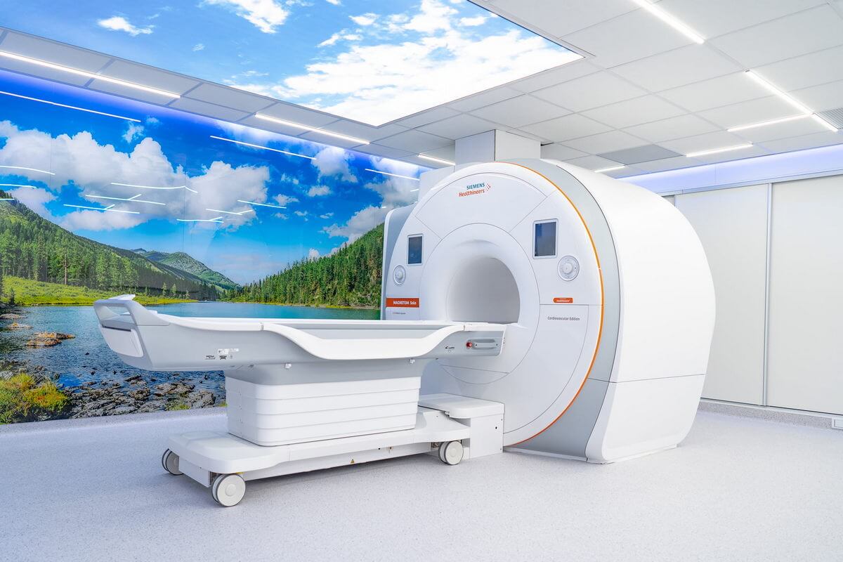
MRI scanners detect extremely faint radio-frequency signals emitted by hydrogen atoms in the patient's body — signals measured in microvolts. Any external electromagnetic energy at or near the scanner's operating frequency that reaches the receive coil will be superimposed on the diagnostic signal, producing artifacts in the final image. This is why every MRI installation requires a Faraday cage — and why even small breaches in that cage can have outsized effects on image quality.
When MRI artifacts appear or worsen over time, the cause is often an electromagnetic interference (EMI) or radio-frequency interference (RFI) source that is either new to the environment, or that is reaching the scanner through a degraded path in the shielding system. Identifying and eliminating these sources is a systematic process that starts with understanding what you're looking for.
How EMI Becomes an Image Artifact
MRI scanners operate at a specific Larmor frequency determined by the static magnetic field strength: 63.87 MHz for 1.5T and 127.74 MHz for 3T systems. The scanner's receive chain is tuned to this frequency and is extraordinarily sensitive — it must be, to detect the weak NMR signals from tissue.
External RF energy that enters the scanner room and reaches the receive coil at or near the Larmor frequency is indistinguishable from the desired signal. The scanner's signal processing interprets this noise as coming from within the patient, encoding it into the image as:
- Zipper artifacts: narrow bright or dark lines running across the image in the frequency-encoding direction. These are the hallmark of a discrete, narrowband RF interference source at or near the Larmor frequency.
- Herringbone (corduroy) patterns: a fine criss-cross pattern overlaid on the image, typically caused by broadband or pulsed EMI that interacts with the phase-encoding gradient.
- Increased noise floor: a general haziness or graininess across the image without a specific pattern, caused by broadband RF energy that raises the overall noise level. This reduces signal-to-noise ratio (SNR) and can make subtle diagnostic findings difficult to detect.
- Ghosting: repeated faint copies of the image shifted in the phase-encoding direction, which can be caused by periodic EMI sources synchronized with the scanner's repetition time.
- Bright spots or streaks: localized signal spikes caused by strong, intermittent interference — such as an arc from faulty electrical equipment or a nearby transmitter keying on and off.
The specific artifact pattern provides the first diagnostic clue. A zipper artifact points to a narrowband source; a general SNR loss suggests broadband contamination; periodic ghosting implies a source with a regular duty cycle.
Common External EMI/RFI Sources
External sources are RF emitters located outside the MRI room whose energy penetrates the Faraday cage through shielding defects. These sources exist in the building and surrounding environment regardless of the MRI installation:
Building Infrastructure
- Variable-frequency drives (VFDs): used in modern HVAC systems, elevators, and pumps. VFDs generate broadband RF noise as a byproduct of their switching operation. If a VFD-controlled motor is on a power circuit that shares the same electrical panel as the MRI room, conducted emissions can enter through the penetration panel power filters.
- LED lighting with switching power supplies: LED drivers can generate RF noise in the MHz range. Fluorescent ballasts — especially aging magnetic ballasts — are another common source.
- Elevators: both the motor drive and the control electronics generate broadband EMI. Elevator shafts running near the MRI suite are a frequent source of intermittent artifacts.
- Uninterruptible power supplies (UPS): the inverter stage in online UPS systems generates switching noise that can be conducted into the MRI room via the power feed.
Nearby Equipment
- Electrosurgical units (ESUs) and cautery equipment: operating rooms adjacent to or above/below the MRI suite can generate intense RF bursts during surgical procedures. ESU frequencies often fall near or within MRI receive bandwidths.
- Physiotherapy diathermy equipment: shortwave (27.12 MHz) and microwave diathermy units are powerful RF emitters that can interfere with MRI if operated in the same facility without adequate separation or shielding.
- Other imaging equipment: CT scanners, X-ray generators, and linear accelerators produce broadband EMI during operation.
External Broadcast and Communication
- FM radio stations: the FM broadcast band (88–108 MHz) overlaps with the 3T Larmor frequency (127.74 MHz) closely enough that harmonics or strong local stations can be detected by inadequately shielded 3T scanners.
- Two-way radios and pagers: hospital staff using handheld radios near the MRI suite generate intermittent, strong RF signals.
- Cellular base stations: cell towers on or near the hospital building produce continuous RF energy in bands that can generate intermodulation products near MRI frequencies.
- Emergency vehicles: ambulance and fire truck radio transmissions from the adjacent parking area or helipad can produce brief, intense RF interference.
Internal Sources (Inside the Faraday Cage)
Not all EMI originates outside the shielded room. Some sources are inside the Faraday cage — and because they're already past the shielding barrier, even low-power emissions can cause significant artifacts:
- Patient monitoring equipment: pulse oximeters, ECG leads, infusion pumps, and ventilators brought into the scanner room can generate RF noise if they are not MRI-compatible (MR Conditional or MR Safe rated). Even MR-compatible equipment can cause artifacts if cables are routed improperly or if devices malfunction.
- LED room lighting: LED drivers inside the shielded room must be RF-quiet. Standard commercial LED fixtures often contain switching regulators that emit noise in the MHz range. MRI-specific LED lighting is designed with filtered drivers to eliminate this.
- In-room speakers and music systems: patient comfort systems that use conventional audio amplifiers can introduce RF noise. MRI-compatible audio systems use filtered or fiber-optic signal paths.
- CCTV cameras: in-room cameras with active electronics (especially wireless cameras, which are prohibited in the MRI room) can generate RF noise. MRI-compatible cameras use shielded housings and filtered power.
- Faulty scanner components: the scanner itself can be a source of artifacts — damaged gradient cables, worn RF coil connectors, or malfunctioning pre-amplifiers can introduce spurious signals. These are typically identified by the scanner manufacturer's service engineer during routine maintenance.
Shielding Defects That Allow EMI Entry
External EMI can only reach the scanner if there is a path through the Faraday cage. When artifacts appear or worsen, the shielding system should be evaluated for these common defects:
- Door seal degradation: the RF shielded door is the most mechanically stressed component of the cage. Worn finger-stock contacts, warped door leaves, or misaligned latches create gaps that leak RF. Door-related leaks often produce artifacts that come and go — worse when the door is not fully compressed, better when it is firmly latched.
- Failed penetration panel filters: a blown Pi-filter is an unfiltered conductor through the cage. Power line filter failures are especially impactful because mains wiring carries conducted EMI from the entire building electrical system.
- Unauthorized penetrations: holes drilled through the cage wall for new cables (bypassing the penetration panel) are one of the most common causes of sudden SE degradation in operating MRI suites.
- Window frame degradation: corrosion or mechanical loosening at the observation window frame bond can create an RF leak. This is less common than door issues but should be checked.
- Panel joint deterioration: corrosion, mechanical stress, or building settlement can degrade the conductive bond at panel joints, particularly at wall-floor and wall-ceiling transitions.
- Waveguide compromises: honeycomb waveguide inserts in HVAC penetrations can become damaged or blocked with debris, altering their RF attenuation characteristics.
The Troubleshooting Process
Step 1: Characterize the Artifact
Document the artifact pattern (zipper, herringbone, diffuse noise, ghosting), its frequency of occurrence (constant, intermittent, time-of-day dependent), and which scan sequences are most affected. Artifacts that appear only during weekday business hours suggest an external source tied to building activity. Artifacts present on phantom scans (no patient) confirm the cause is environmental rather than patient-related.
Step 2: RF Noise Survey
Use a spectrum analyzer with a near-field probe to measure RF emissions at multiple points around the shielded room — outside the cage, inside the cage, at the door perimeter, at the penetration panel, and at each waveguide entry. Comparing inside-vs-outside measurements quantifies the room's actual SE and identifies localized leak points.
Step 3: Systematic Elimination
Turn off building systems one at a time (HVAC, elevators, lighting circuits, nearby medical equipment) while monitoring the noise floor inside the MRI room. When the artifact disappears after switching off a specific system, the source is identified. For intermittent sources, this may require extended monitoring over hours or days.
Step 4: Full SE Test
If the noise survey reveals SE degradation compared to the room's baseline (commissioning) SE data, a full IEEE 299 test identifies exactly which surfaces, joints, or components have lost performance. This directs the repair effort to the specific defect.
Step 5: Remediation
Depending on findings, remediation may include: replacing worn door seals, replacing failed penetration panel filters, sealing unauthorized penetrations, re-bonding window frames, upgrading power filters to address newly identified conducted EMI sources, or relocating external equipment. In some cases, a shielding retrofit may be warranted if the cage has degraded beyond simple repair.
Prevention Strategies
Preventing EMI problems is far less costly and disruptive than troubleshooting them after they appear:
- Annual SE spot checks: measure SE at the door, window, penetration panel, and representative wall/ceiling points every year. Compare results to the baseline commissioning data to detect gradual degradation before it reaches the artifact threshold.
- Pre-installation EMI site survey: before installing a new MRI scanner (or before building the shielded room), conduct an RF noise survey of the proposed site to identify existing EMI sources. This data informs the shielding material selection, SE specification, and penetration panel filter design.
- Change control: establish a policy requiring that any modification to the MRI suite — new cables, new equipment, building renovations nearby — be reviewed by the MRI physicist or shielding contractor to assess potential EMI impact. No penetration of the Faraday cage should occur without going through the penetration panel.
- MR-compatible equipment only: all equipment brought into the scanner room must be rated MR Safe or MR Conditional, and its RF emission characteristics should be verified. Even MR-compatible equipment should be tested in place to confirm it does not introduce artifacts.
- Facility coordination: maintain communication with the facility engineering team so that new equipment installations (VFDs, generators, cell repeaters) near the MRI suite are evaluated for potential EMI impact before they are commissioned.
- Maintain ACR Zone controls: the zone access system limits what equipment and personnel enter the scanner room, which is also an effective EMI prevention measure.
Frequently Asked Questions
What does EMI interference look like on an MRI image?
The most common pattern is a "zipper artifact" — a narrow bright or dark line running across the image in the frequency-encoding direction, caused by a discrete RF source near the Larmor frequency. Other patterns include herringbone (criss-cross) patterns from broadband or pulsed EMI, general image graininess from elevated noise floor, and ghosting from periodic interference sources. The specific pattern helps identify the type of EMI source.
Can a cell phone cause MRI image artifacts?
Yes. Cell phones are powerful RF transmitters that emit energy in bands that can couple into the MRI receive chain, either directly (if brought into the scanner room) or through shielding defects (if used nearby). Cell phones should never be brought into the MRI scanner room, and their use should be restricted in the immediate vicinity of the shielded room per ACR zone access controls.
How do you find the source of RF interference in an MRI room?
The standard approach uses a spectrum analyzer with a near-field probe to measure RF emissions at multiple points inside and outside the shielded room, identifying leak paths and characterizing the interference frequency. Combined with systematic elimination (turning off building systems one at a time while monitoring the MRI noise floor), this process isolates both the source and the entry path.
Can elevator operation cause MRI artifacts?
Yes. Elevator motor drives and control electronics generate broadband EMI, and the elevator shaft acts as a waveguide that can channel this energy. Elevators are a common source of intermittent MRI artifacts, especially in facilities where the elevator shaft runs near or adjacent to the MRI suite. The artifacts typically appear as brief bursts of noise during elevator movement.
How often should MRI room shielding be tested for EMI leaks?
Annual shielding effectiveness (SE) spot checks at key locations (door, window, penetration panel, representative wall points) are the industry standard. A full IEEE 299 SE test should be performed whenever artifacts suggest shielding degradation, after any modification to the Faraday cage, or as part of a pre-upgrade assessment before a new scanner installation.