Introduction
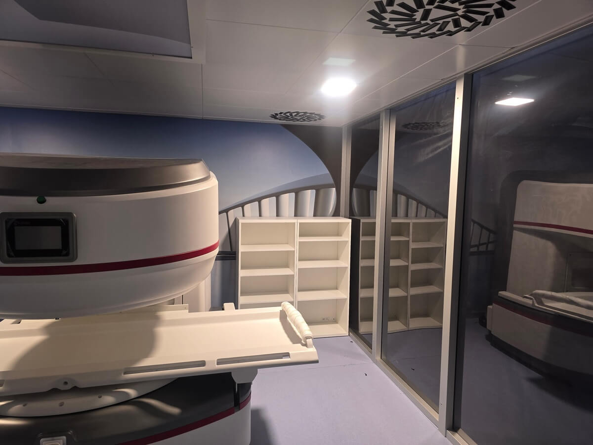
Superconducting MRI magnets operate at temperatures near absolute zero, maintained by a bath or closed-loop system of liquid helium. A magnet quench occurs when a section of the superconducting coil loses its superconducting state and becomes resistive, causing the stored magnetic energy to convert rapidly into heat. This boils off the liquid helium in seconds, producing a massive volume of gaseous helium that must be safely vented outside the building.
The quench pipe (also called a quench vent or cryogen exhaust) is the engineered pathway for this emergency helium release. If the quench pipe is undersized, obstructed, or improperly designed, helium can backflow into the MRI scanner room — displacing breathable oxygen and creating a life-threatening asphyxiation hazard. Combined with emergency room ventilation and oxygen depletion monitoring, the quench system is one of the most safety-critical elements of any MRI suite design.
This guide covers quench pipe sizing, routing, and materials; emergency ventilation requirements; oxygen monitoring; and how the quench system integrates with the Faraday cage and ACR safety zone design.
What Happens During a Magnet Quench
The Physics
A superconducting MRI magnet stores enormous energy in its magnetic field — a 1.5T clinical scanner stores roughly 3–5 MJ, while a 3T system may store 15–30 MJ or more. During a quench, this energy converts to heat within seconds. The heat boils the liquid helium (boiling point: −269°C / 4.2 K), and the resulting gas expands by a factor of approximately 750:1 as it warms to room temperature.
A typical 1.5T scanner contains 1,500–2,000 liters of liquid helium. A full quench can produce over 1,000 cubic meters of gaseous helium at atmospheric pressure — roughly enough to fill a large MRI scanner room 5–8 times over. This gas must exit the room in under 60 seconds to prevent dangerous pressure buildup and oxygen displacement.
The Danger
Helium is non-toxic but is an asphyxiant. Because it is lighter than air, it rises and accumulates at the ceiling, pushing breathable air downward. In a sealed room, helium can displace enough oxygen to cause unconsciousness within minutes and death within 10–15 minutes. The cold gas can also cause frostbite on contact and may damage equipment. If the room overpressurizes (due to a blocked or undersized quench pipe), the RF shielded door and observation window can be blown open or structurally damaged.
Quench Pipe Design Requirements
Pipe Sizing
The quench pipe diameter is specified by the MRI scanner manufacturer based on the magnet's helium inventory, quench rate, and maximum allowable back-pressure. Typical requirements range from 200 mm (8 inches) for 1.5T systems to 300 mm (12 inches) or larger for 3T and high-field systems. Undersizing the pipe is the single most dangerous design error — it restricts helium flow, causing pressure to build inside the scanner room.
Routing
The quench pipe runs from the scanner's cryostat (usually exiting the top of the magnet) through the ceiling, up through the building structure, and terminates outside the building at a safe discharge point. Key routing principles include:
- Shortest path possible: every meter of pipe adds flow resistance. The fewer bends and the shorter the total length, the lower the back-pressure during a quench.
- Minimize bends: each 90° elbow adds significant flow resistance. Use 45° bends or long-radius sweeps where changes in direction are unavoidable. Most manufacturer specifications limit the total number and angle of bends.
- Continuous upward slope: the pipe should slope upward continuously from the magnet to the discharge point to prevent condensate (water from humid air or cold helium condensation) from pooling and partially blocking the pipe.
- No valves or dampers: the quench pipe must never be fitted with any valve, damper, cap, or obstruction that could restrict flow. Rain caps at the exterior discharge point must be the gravity-opening type that cannot seal shut.
Materials
The pipe must withstand the extreme thermal shock of cryogenic helium (initially near −269°C) followed by rapid warming. Stainless steel (typically 304 or 316L) is the standard material. Carbon steel and PVC are not acceptable — carbon steel becomes brittle at cryogenic temperatures, and PVC shatters. All joints must be welded or flanged with cryogenic-rated gaskets; threaded connections are not reliable under thermal cycling.
Discharge Point
The pipe terminates outside the building at a location where the discharged helium cannot re-enter the building or endanger people. Requirements typically include:
- Minimum 3 meters (10 feet) above any occupied area, walkway, or air intake
- Directed away from building air intakes, windows, and doorways
- Not in an enclosed courtyard or recessed area where helium could accumulate
- Protected with a rain cap that opens freely under quench pressure
Rupture Discs and Pressure Relief
A rupture disc (burst disc) is a thin metallic membrane installed in the quench pipe system that provides a secondary pressure relief path. If the primary quench pipe becomes obstructed (ice blockage, debris, improper modifications), the rupture disc bursts at a predetermined pressure to vent helium into an alternative path — typically directly into the scanner room or an adjacent space.
This is a worst-case safety mechanism: venting helium into the room is dangerous, but it prevents the cryostat from rupturing under pressure (which could cause catastrophic magnet failure and shrapnel). The rupture disc is sized and located per the scanner manufacturer's specification and must never be modified, relocated, or obstructed.
Some installations include a secondary rupture disc that vents to the outdoors through an alternative pipe route, providing redundant exterior venting before room release is necessary.
Emergency Room Ventilation
Even with a properly functioning quench pipe, some helium may escape into the scanner room — through the rupture disc path, through imperfect seals at the magnet-to-pipe connection, or during a slow boil-off event that doesn't fully pressurize the quench pipe. The MRI room's emergency ventilation system is the second line of defense.
Design Requirements
Emergency ventilation for the MRI scanner room (distinct from the normal HVAC system) must provide rapid air exchange to dilute and remove helium. Key specifications per NFPA 99 and ASHRAE guidelines include:
- Exhaust rate: sufficient to provide a minimum of one complete room air change within 60–120 seconds. For a typical MRI room (50–80 m³), this translates to exhaust fans capable of 1,500–5,000 CFM depending on room volume and local code requirements.
- Exhaust at ceiling level: because helium rises, the emergency exhaust grille must be located at or near the ceiling — not at floor level like normal HVAC returns.
- Automatic activation: the emergency exhaust is triggered automatically by the oxygen depletion sensor system (see below). Manual activation switches should also be provided at the MRI room entrance and in the control room.
- Makeup air: the emergency exhaust must be paired with a makeup air supply (from the corridor or adjacent space) to prevent the room from going into negative pressure, which would resist door opening and impede evacuation.
- Independent circuit: the emergency exhaust fan must be on a dedicated electrical circuit with emergency/backup power so it operates during a building power failure.
Integration with the Faraday Cage
The emergency exhaust duct penetrates the Faraday cage, which means it must pass through a waveguide to maintain shielding effectiveness. The waveguide dimensions must be large enough to pass the required airflow while still providing RF attenuation. Honeycomb waveguide inserts are typically used — the same technology used for the normal HVAC penetrations, but sized for the higher emergency exhaust airflow rate.
Oxygen Depletion Monitoring
Oxygen depletion sensors (ODS) are the early warning system that detects helium accumulation in the MRI room before oxygen levels become dangerous. NFPA 99 and ACR guidelines require ODS in all MRI scanner rooms containing superconducting magnets.
Sensor Placement
Sensors are mounted near the ceiling (where helium accumulates first) and at head height (to detect oxygen displacement in the breathing zone). Most installations use a minimum of two sensors: one ceiling-mounted and one at approximately 1.5 m (5 feet) above the floor. Larger rooms or rooms with complex geometries may require additional sensors.
Alarm Thresholds
Normal atmospheric oxygen concentration is 20.9%. Standard alarm thresholds are:
- First alarm (caution): 19.5% O₂ — activates a visual and audible warning. Staff should prepare to evacuate and investigate the cause.
- Second alarm (danger): 18.0% O₂ — activates emergency exhaust ventilation automatically, triggers building alarm, and requires immediate evacuation of the MRI room. Oxygen levels below 16% cause impaired judgment and coordination; below 10% causes unconsciousness within minutes.
Alarm Annunciation
ODS alarms must be visible and audible both inside the MRI room and at the Zone III control area. Alarm panels are typically located at the MRI room entrance (Zone III/IV boundary) and in the control room. Integration with the building fire alarm or building management system may be required by local code.
Maintenance
Oxygen sensors require regular calibration — typically every 6–12 months depending on the sensor type. Electrochemical sensors have a finite lifespan (typically 2–3 years) and must be replaced before their rated expiration. Sensor calibration and replacement must be documented in the MRI suite safety log.
US Regulatory Framework
Quench pipe and emergency ventilation design in the United States is governed by several overlapping standards:
- NFPA 99 (Health Care Facilities Code): Chapter 12 addresses MRI-specific requirements including quench venting, oxygen monitoring, and emergency ventilation. This is the primary code referenced by most state and local building authorities.
- ASHRAE Handbook — HVAC Applications: provides guidance on ventilation rates and exhaust system design for MRI suites, including emergency exhaust sizing calculations.
- ACR Guidance Document on MR Safe Practices: while not a building code, the ACR guidance is referenced by accreditation bodies (including The Joint Commission) and establishes best-practice requirements for oxygen monitoring and emergency procedures.
- MRI manufacturer site planning documents: each scanner manufacturer (Siemens, GE, Philips, Canon) publishes detailed site planning requirements that include quench pipe specifications, maximum allowable back-pressure, pipe diameter, and routing constraints specific to their magnet design.
- State and local building codes: requirements vary by jurisdiction. Some states (California, New York, Florida) have additional requirements beyond NFPA 99 for medical gas systems and emergency ventilation in healthcare facilities.
The quench pipe design must satisfy all applicable codes and the scanner manufacturer's specifications — whichever is most restrictive governs. During the design phase, the mechanical engineer, shielding contractor, and MRI vendor should collaborate to ensure the quench system is integrated with the Faraday cage design and meets every requirement.
Frequently Asked Questions
What is an MRI quench and why is it dangerous?
A quench occurs when a superconducting MRI magnet loses its superconducting state, causing rapid boil-off of liquid helium. The helium expands roughly 750:1 as it warms to room temperature, producing over 1,000 cubic meters of gas from a typical 1.5T scanner. Helium is non-toxic but displaces breathable oxygen — if it accumulates in the scanner room, it can cause unconsciousness and death by asphyxiation within minutes.
How big does an MRI quench pipe need to be?
Quench pipe diameter is specified by the MRI scanner manufacturer, typically 200 mm (8 inches) for 1.5T systems and 300 mm (12 inches) or larger for 3T systems. The pipe must be large enough to vent the full helium volume without exceeding the maximum allowable back-pressure at the magnet. Never use a smaller pipe than specified — undersizing is the most dangerous quench system design error.
Does the quench pipe affect MRI room shielding?
Yes — the quench pipe passes through the Faraday cage, creating a potential RF leak path. The pipe penetration must be designed as a waveguide with sufficient length-to-diameter ratio to attenuate RF at the MRI operating frequency. The shielding contractor and mechanical engineer must coordinate to ensure the quench pipe penetration maintains the room's shielding effectiveness specification.
What are oxygen depletion sensors and where are they required?
Oxygen depletion sensors (ODS) monitor the oxygen concentration in the MRI room and trigger alarms if helium displaces breathable air. They are required by NFPA 99 and ACR guidelines in all rooms containing superconducting MRI magnets. Sensors are typically mounted near the ceiling and at head height, with alarms at 19.5% O₂ (caution) and 18.0% O₂ (danger/evacuate).
Can a quench damage the MRI room or Faraday cage?
If the quench pipe is obstructed or undersized, the rapid pressure buildup can blow open the RF shielded door, damage the observation window, and crack interior finishes. The Faraday cage panels themselves are typically robust enough to withstand moderate overpressure, but door seals and window frames are vulnerable. A properly designed and maintained quench pipe prevents this by venting helium outside the building before pressure builds.